


Persistent pain in the groin or hip is often dismissed as a muscle strain or arthritis. However, in some patients, the underlying cause is Avascular Necrosis (AVN) of the hip, a condition in which the blood supply to the femoral head is reduced or completely interrupted. Without adequate blood flow, the bone gradually weakens and may collapse, leading to severe pain, stiffness and loss of hip function.
AVN can affect adults of different age groups and often progresses silently during its early stages. Some people experience only mild discomfort initially, while others notice increasing difficulty walking, climbing stairs or performing everyday activities. Because the damage develops over time, delaying treatment can reduce the chances of preserving the natural hip joint.
The good news is that AVN can often be managed successfully when diagnosed early. Depending on the stage of the disease, treatment may include medication, activity modification, physiotherapy, hip-preserving surgery such as core decompression, or total hip replacement when the joint has sustained significant damage. At Mangal Anand Hospital, our orthopaedic surgeons evaluate every patient individually to recommend the most appropriate treatment based on the stage of AVN, symptoms, lifestyle and overall joint health.

Avascular Necrosis (AVN), also called osteonecrosis of the hip, is a condition in which the blood supply to the femoral head, the ball-shaped upper end of the thigh bone, becomes reduced or completely blocked. Since bone tissue depends on a constant blood supply to remain healthy, interruption of circulation causes the bone cells to die gradually.
In the early stages, the shape of the hip joint may remain normal even though damage has already begun inside the bone. As the condition progresses, the weakened femoral head may collapse, causing the smooth cartilage covering the joint to wear out. Once this happens, patients often develop persistent pain, stiffness and difficulty walking.
Although AVN can affect different bones in the body, the hip is the most commonly involved joint because it relies on a delicate blood supply. The condition may affect one hip or, in some patients, both hips.
Early diagnosis is important because treatment options are generally more effective before the femoral head collapses.
The symptoms of AVN usually develop gradually. Many patients experience mild discomfort initially and may not realise that the condition is progressing. Common symptoms include:
As the disease progresses and the femoral head begins to collapse, everyday activities such as sitting, getting out of a chair or wearing footwear may become increasingly difficult
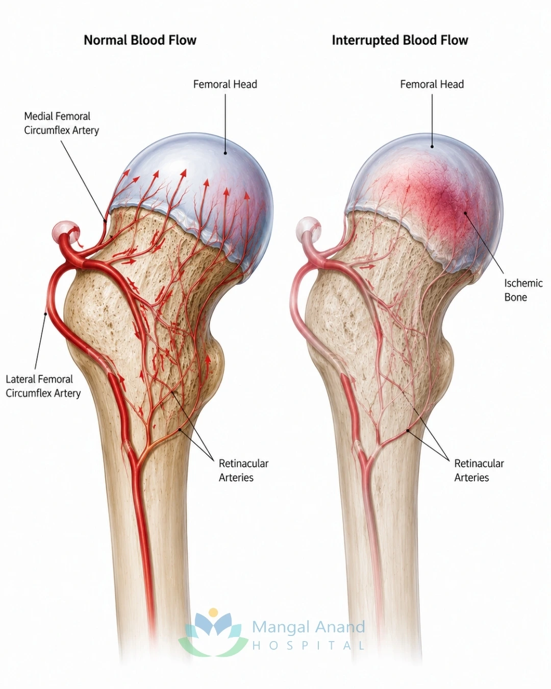
AVN develops when the blood supply to the femoral head is interrupted. In some patients, the exact cause cannot be identified. This is known as idiopathic AVN.
However, several conditions are known to increase the risk of developing AVN.
These include:
Not everyone with these risk factors develops AVN, but individuals with persistent hip pain should undergo evaluation to rule out the condition.
Hip replacement surgery at Mangal Anand Hospital is performed by an experienced team of orthopaedic surgeons with expertise in joint replacement, complex trauma, sports injuries, and reconstructive surgery.
Dr. Milind Sawant is the primary consultant for hip replacement surgery at Mangal Anand Hospital. With orthopaedic training in both India and the United Kingdom, he brings extensive experience in joint replacement and reconstructive orthopaedic surgery.
His qualifications include FRCS (Orthopaedics), FRCS (Edinburgh), FRCS (Glasgow), DNB Orthopaedics, FCPS, MS Orthopaedics and D.Orth. He has completed advanced fellowships in Knee & Sports Medicine in England, Knee Surgery in Australia, and Revision Knee Replacement in Germany and the USA.
In addition to clinical practice, Dr. Sawant has served as faculty for programmes that train orthopaedic surgeons in arthroscopy, knee replacement and fracture management. He has authored numerous peer-reviewed publications and presented research at national and international orthopaedic meetings.

Consultant Orthopaedic Surgeon
Qualifications
Special Interest
Dr. Pradip Nemade has been managing complex orthopaedic conditions since 2009, with a special focus on hip disorders, joint replacement and complex trauma. His clinical approach emphasises accurate diagnosis, evidence-based treatment and restoring mobility while helping patients make informed decisions about their care.

Qualifications
Special Interest
Dr. Nikhil Gokhale graduated from Seth G.S. Medical College and K.E.M. Hospital, Mumbai, and completed advanced fellowship training in the United Kingdom. His training includes the Exeter Knee Fellowship, Shoulder Fellowship at Royal Bournemouth Hospital and a specialised fellowship in Revision Joint Replacement Surgery at the Royal National Orthopaedic Hospital,

AVN is more commonly seen in adults between 30 and 60 years of age, although it can occur earlier depending on the underlying cause.You may have a higher risk if you:
Recognising these risk factors allows earlier investigation before permanent joint damage occurs.
The goal of AVN treatment is to relieve pain, preserve the natural hip joint whenever possible and help you return to an active lifestyle. The right treatment depends on how much damage has occurred to the femoral head, your symptoms, age, activity level and overall health.
If AVN is diagnosed in its early stages, surgery may not always be necessary. However, once the femoral head collapses and arthritis develops, hip replacement often provides the best long-term outcome.
Yes, some patients with early-stage AVN can initially be managed without surgery. While non-surgical treatment cannot restore dead bone, it may help relieve symptoms, reduce stress on the hip and slow disease progression in selected patients.
Non-surgical treatment is generally considered when:
Regular follow-up is important because AVN can continue to progress even when symptoms temporarily improve.
Medicines cannot reverse AVN, but they can help manage pain and inflammation.
Your orthopaedic surgeon may recommend:
Patients should avoid self-medicating for prolonged periods, as persistent pain may indicate worsening bone damage that requires further evaluation.
Reducing excessive stress on the affected hip may help relieve symptoms during the early stages of AVN.
Your doctor may advise you to:
These measures aim to reduce pain while treatment is planned. They do not cure AVN
Physiotherapy plays an important role in maintaining mobility and improving muscle strength around the hip. A physiotherapy programme may include:
Physiotherapy helps improve function but cannot stop the progression of AVN once the blood supply to the bone has been lost.
Core decompression is one of the most commonly performed procedures for early-stage AVN before the femoral head collapses. During the procedure, the surgeon creates one or more small channels within the affected bone to reduce pressure and improve blood flow. This may help relieve pain and encourage healing in carefully selected patients.
Core decompression is generally considered when:
The success of the procedure depends largely on how early the condition is diagnosed. Patients with advanced AVN are less likely to benefit from this procedure.
In selected patients, bone grafting may be performed along with core decompression to provide additional structural support and encourage bone healing.The graft may be taken from another part of the patient's body or obtained from a donor, depending on the surgical plan.Bone grafting is not required for every patient and is recommended only in specific clinical situations.
Stem cell therapy has been studied as an additional treatment for selected patients with early AVN, often in combination with core decompression.While some studies have shown encouraging results, research is still evolving. Stem cell therapy is not considered a standard treatment for every patient with AVN.
Your orthopaedic surgeon will discuss whether this option is appropriate based on your condition and the latest available evidence.
When Is Hip Replacement Required?
Hip replacement surgery is usually recommended when AVN has caused significant collapse of the femoral head or when arthritis has developed within the joint. In these situations, hip replacement surgery helps relieve pain, restore movement and improve quality of life when hip-preserving procedures are no longer likely to be effective.
Hip replacement may be recommended if you:
Modern hip replacement surgery allows many patients to return to walking comfortably and resume their normal daily activities after rehabilitation.
AVN is generally a progressive condition. Without appropriate treatment, the blood-deprived bone continues to weaken. As the disease advances, patients may experience:
Seeking treatment during the early stages offers the best opportunity to preserve the natural hip joint and maintain long-term function.
Choosing the right hospital for AVN treatment is important because treatment decisions vary depending on the stage of the disease. Early-stage AVN may be managed with hip-preserving procedures, while advanced cases often require hip replacement surgery. At Mangal Anand Hospital, every patient undergoes a thorough evaluation to determine the most appropriate treatment based on their condition, symptoms and lifestyle.
Our orthopaedic team has experience in managing a wide range of hip disorders, from early AVN to advanced joint damage. Treatment plans are individualised with the aim of relieving pain, improving mobility and helping patients return to their daily activities as safely as possible.
Patients undergoing treatment for AVN at Mangal Anand Hospital have access to:
Our team works together to ensure continuity of care from diagnosis through rehabilitation.
AVN progresses gradually, and treatment depends largely on the stage of the disease. Understanding the stage helps you know whether the hip joint can still be preserved or whether joint replacement is likely to provide better long-term results.
The blood supply to the femoral head has been affected, but the bone has not collapsed. Many patients experience pain during walking or physical activity, while X-rays may still appear normal. MRI usually detects the disease at this stage.Patients diagnosed early may benefit from non-surgical treatment or hip-preserving procedures such as core decompression, depending on the extent of bone involvement.
As AVN progresses, the affected bone becomes weaker and early structural changes may develop. Pain usually becomes more frequent, and daily activities such as climbing stairs or walking long distances may become uncomfortable. Treatment at this stage depends on the amount of bone damage, the patient's symptoms and overall hip function. While some patients may still be candidates for hip-preserving surgery, others may require joint replacement.
In advanced AVN, the femoral head loses its normal shape and eventually collapses. The damaged joint surface leads to arthritis, causing persistent pain, stiffness and significant restriction of movement.
For many patients with advanced AVN, total hip replacement provides the most reliable long-term pain relief and improvement in mobility. Early diagnosis offers the best opportunity to preserve the natural hip joint before permanent damage occurs.
AVN (Avascular Necrosis) is a condition in which the blood supply to the femoral head is reduced or interrupted. Over time, the affected bone weakens and may collapse, leading to pain and arthritis of the hip.
Early symptoms usually include pain in the groin, hip or thigh, especially while walking or climbing stairs. Some patients may also experience stiffness or a limp.
AVN does not usually heal on its own. Early-stage disease may be managed without surgery in selected patients, but regular monitoring is essential because the condition can progress over time.
No. Patients diagnosed during the early stages may be suitable for hip-preserving treatments. Hip replacement is generally recommended when the femoral head has collapsed or the hip joint has developed advanced arthritis.
Core decompression is a surgical procedure performed during the early stages of AVN. Small channels are created within the affected bone to reduce pressure and encourage improved blood flow.
MRI is the most sensitive investigation for detecting AVN and is often able to identify the condition before changes become visible on an X-ray.
Yes. AVN may affect one or both hips. If both hips are involved, one side is often more painful than the other.
Common causes include long-term steroid use, excessive alcohol consumption, hip fractures or dislocations, sickle cell disease and certain autoimmune disorders. In some patients, no specific cause is identified.
Walking is generally safe within the limits advised by your orthopaedic surgeon. High-impact activities may need to be avoided to reduce stress on the affected hip.
Yes. Unlike hip arthritis, AVN often affects adults between 30 and 60 years of age, although it can occur earlier depending on the underlying cause.
Recovery varies between individuals. Most patients gradually return to walking and routine daily activities with physiotherapy and rehabilitation over the weeks following surgery.
The treated hip is managed according to the procedure performed. However, patients with ongoing risk factors should continue regular follow-up, and those with AVN in one hip may require monitoring of the other hip.
AVN should be evaluated by an orthopaedic surgeon experienced in the diagnosis and treatment of hip disorders, including hip-preserving procedures and hip replacement surgery.
Hip Replacement Surgery
Revision Hip Replacement
Orthopaedic Trauma Care
Osteoporosis Management
Physiotherapy and Rehabilitation
Joint Replacement Surgery
Dr. Milind Sawant – Profile
Dr. Pradip Nemade – Profile